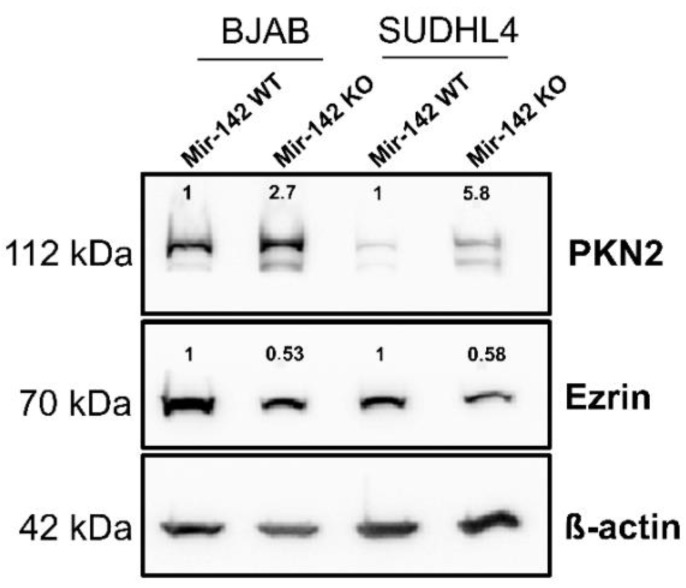
Figure 11.
Western blot analysis: Extracts of BJAB-wild-type and SUDHL4-wild-type (“WT”) cells and the corresponding miR-142 knockout (“KO”) cells were separated on a 10% polyacrylamide gel, transferred to a nitrocellulose membrane and incubated with antibodies against PKN2, Ezrin and β-actin as a loading control. The membranes were incubated with the appropriate secondary antibodies coupled to horseradish peroxidase. Bound secondary antibodies were visualized by ECL. The bands were quantified using Image Lab 6.0.1. The mean value of two separate experiments is shown above the bands. The wild-type value was set to 1. The uncropped blots are shown in Figure S4.

