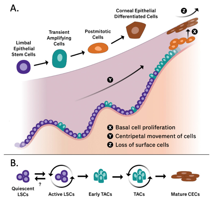
Figure 2.
(A) Graphical representation of the limbal stem cell niche containing all cell states that have been implicated in corneal epithelium regeneration (LSCs, TACs, postmitotic cells, and corneal epithelial differentiated cells). The X, Y, Z hypothesis published by Thoft and Friend in 1983 [32] is also illustrated, including the three phenomena that allow the corneal epithelial cell mass to remain constant. X: proliferation of basal epithelial cells; Y: contribution to the cell mass by centripetal movement of peripheral cells; Z: epithelial cell loss or constant desquamation from the surface. (B) A graphical representation of the differentiation of quiescent limbal stem cells into mature corneal epithelial cells. While the triggers that drive quiescent LSCs into an active differentiating phenotype are well documented, further exploration in reversing this process is warranted. Adapted from [33,34].

