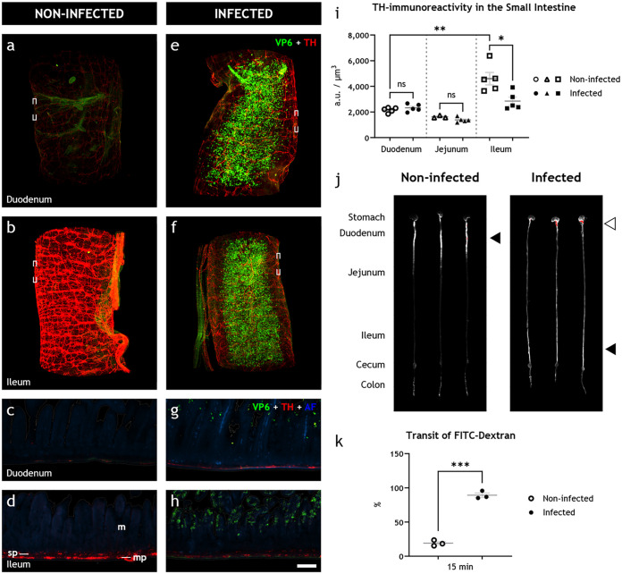
FIG 2.
Rotavirus infection simultaneously leads to increased intestinal transit and downregulation of tyrosine hydroxylase in sympathetic noradrenergic neurons of the autonomic nervous system. (a to h) Light-sheet micrograph stacks (a, b, e, and f) and single optical slice (c, d, g, and h) of infected (right) and noninfected (left) duodenum (a, c, e, and g) and ileum (b, d, f, and h) stained for rotavirus VP6 (green) and TH (red), used as a marker for detecting sympathetic axons. Tissue was visualized with autofluorescence (AF; blue). Micrographs in panels c, d, g, and h correspond to boxed regions in panels a, b, e, and f. Note the reduced level of TH immunoreactivity in infected (f, h) versus noninfected (b, d) ileum. (i) Quantification of TH immunoreactivity, statistically analyzed with two-tailed unpaired (infected versus noninfected) and paired (duodenum versus ileum) t tests, showed no significant difference in the duodenum (ns; P = 0.3236) or jejunum (ns; P = 0.0934) of infected and noninfected animals, a significant increase in the ileum compared to the duodenum of noninfected animals (**, P = 0.0066), and a significant decrease in the ileum of infected compared to noninfected animals (*, P = 0.0157). (j) UV spectrophotometry of the gastrointestinal tracts of noninfected and 16 hpi infant mice 15 min after FITC-dextran treatment. The average travel distance is marked with a black triangle; FITC-dextran remnants in the stomach are marked with a white triangle. (k) Transit of FITC-dextran relative to the entire length of the intestine statistically analyzed with the two-tailed unpaired t test with Welch’s correction showed a significant increase (***, P = 0.0001) in the intestinal motility of the infected animals. m, mucosa; mp, myenteric plexus; sp, submucosal plexus. Scale bar shown in panel h represents 100 μm for panels e to h.

