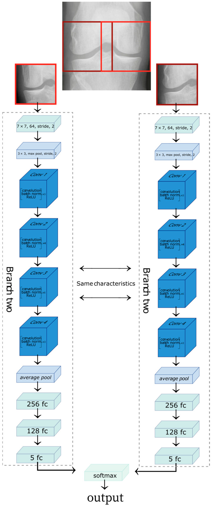
Figure 2.
Representation of the Siamese network architecture. The lateral side of the knee X-ray image feeds one branch of the network, and the medial side of the knee X-ray image provides the second branch of the network. The figure was adapted from [28].

