Figure 3.
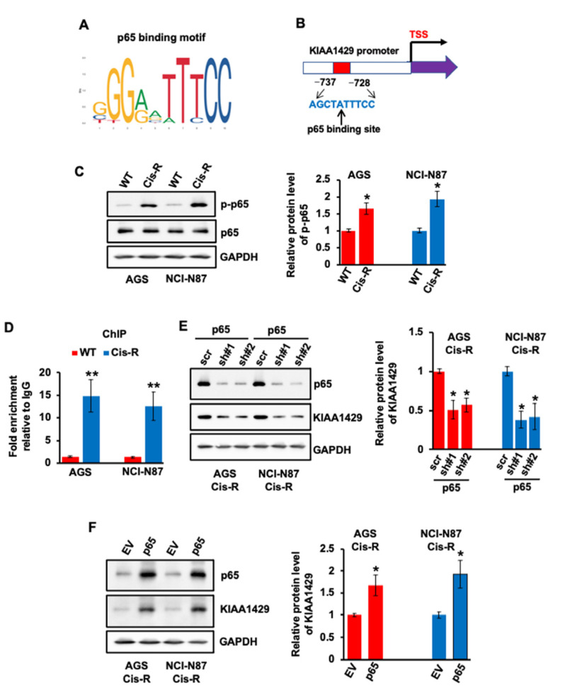
Effect of p65 on regulating KIAA1429 expression. (A) The canonical binding motif of p65. (B) Schematic diagram of KIAA1429 promoter region con-taining one putative p65 binding sites. (C) The phosphorylation of p65 and total p65 levels in AGS Cis-R or NCI-N87 Cis-R and their parental wild type cells was assessed by Western blotting and densitometric quantification. (D) ChIP assay indicated an increase of p65 binding to KIAA1429 promoter in AGS Cis-R or NCI-N87 Cis-R compared with their parental wild type cells. (E) The protein level of KIAA1429 was assessed by Western blotting and densitometric quantification after p65 depletion. (F) The protein level of KIAA1429 was assessed by Western blotting and densitometric quantification after p65 overexpression. * p < 0.05, ** p < 0.01.

