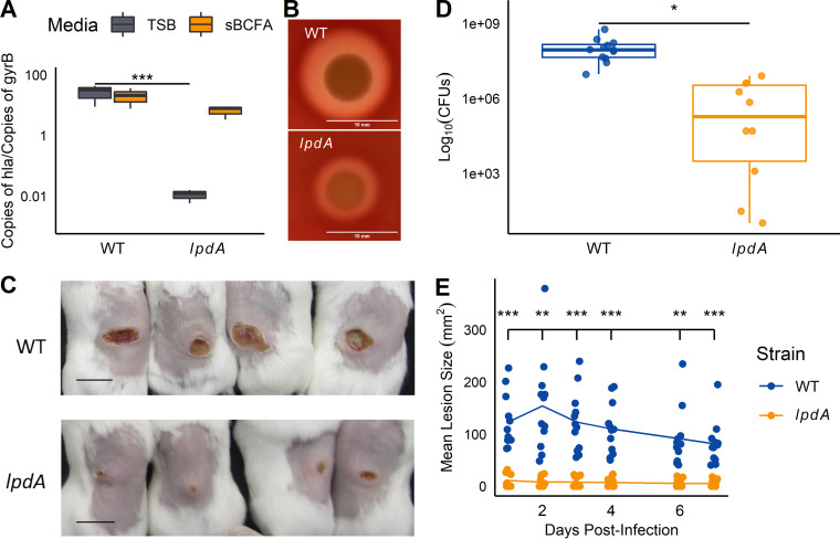
FIG 9.
Hemolysin production and dermonecrosis are reduced in the lpdA mutant. (A) hla transcript levels were quantified in wild-type and lpdA cells grown for 5 h in TSB with or without 0.5 mM sBCFAs using RT-qPCR as described in Materials and Methods. Transcript levels are normalized by gyrase B (gyrB). n = 3. (B) lpdA mutants show reduced hemolysis on sheep blood agar plates. Representative image from 3 trials; bar = 10 mm. (C to E) Mice (n = 12) were infected subcutaneously with wild-type or lpdA S. aureus, and dermonecrotic lesions were measured for 7 days postinfection. (C) Images of representative skin lesions on day 7. Bar, 1 cm. (D) CFU were quantified 7 days after infection. (E) Mean lesion size over 7 days postinfection. *, P < 0.05; **, P < 0.01; ***, P < 0.001, ANOVA with Tukey post hoc test (A), Student’s t test (D), or ANOVA with repeated measures (E).

