FIG 3.
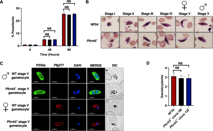
Pfcrk5− asexual stages grow normally and undergo gametocytogenesis. (A) Ring stage synchronous cultures for WT and two clones of Pfcrk5− (clone 5B and 12F) were plated to measure parasite growth over the course of 2 erythrocytic cycles. Total parasitemia was determined by counting the parasites from Giemsa-stained thin blood smears. Data were averaged from three biological replicates and presented as the mean ± standard deviation (SD). ns, not significant unpaired two-tailed Student's t test. (B) Ring stage synchronous cultures for WT and 2 different clones of Pfcrk5− (clone 5B and 12F) were tested for their potential to form gametocytes. Light microscopy of Giemsa-stained smears showing development of WT PfNF54 and Pfcrk5− gametocytes and the 5 (I-V) distinct morphological stages. 1,000×magnifcation. Symbols for female and male gametocytes are shown on top of stage V gametocytes. (C) IFAs were performed on WT PfNF54 and Pfcrk5− mature stage V gametocytes thin culture smears using anti-PfP230p antisera, a marker for stage V male gametocytes (in green), in combination with anti-Pfg377 antisera, a marker for female gametocytes (in red). Representative images are shown. The parasite DNA was visualized with DAPI (blue). Scale bar = 5 μm. Merge I- merged image for red and green panels. Merge II- merged image for red, green, and DAPI (blue) channel. DIC, differential interference contrast. DAPI, 4′,6-diamidino-2-phenylindole. Symbols for male and female gametocytes are shown on left side of the image panels. (D) Gametocytemia was measured on day 15 using thin Giemsa-stained smears. Data were averaged from 3 biological replicates and presented as the mean ± standard deviation (SD). NS, Not significant.

