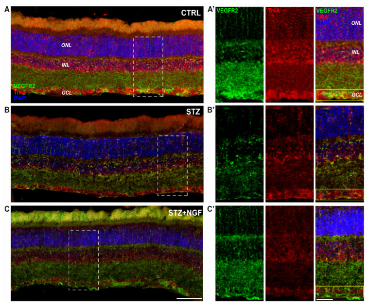
Figure 6.
(A–C). Distribution of TrkA and VEGFR2 in the retinal layers of healthy and diabetic rat retina, with or without topical NGF administration. (A–C) The immunostaining of rat retinal sections showed the different localization of TrkA (red) and VEGFR2 (green), distributed through the retinal layers: ONL, INL, and GCL. Cell nuclei were labeled by DAPI staining (blue). (A’–C’) The high magnification reported the separated channels of TrkA/VEGFR2 staining in the retinal layers, focusing on the GCL. Islets corresponding to the scale bar: 100 µm, islet: 30 µm. CTRL, healthy rats; STZ, streptozotocin; STZ+NGF, streptozotocin+nerve growth factor eye drops.

