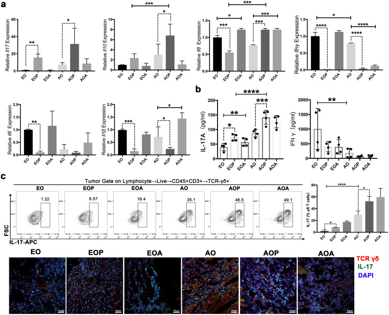
FIG 3.
In the development of OSCC with periodontitis, IL-17+ γδ T cells are activated. (a) The mRNA expression of Il17, Il10, Il18, Ifnγ, Il6, and Il1β in the tumor tissues was detected via qRT-PCR, and expression was normalized to Gapdh. (b) IL-17A and IFN-γ levels in serum were measured by ELISA. (c) IL-17 expression in γδ T cells from OSCC tissues was examined by flow cytometry. Representative plots are shown, and the frequencies of IL-17+ γδ T cells were quantified. Representative confocal immunofluorescence images of OSCC tissues are shown. Red, TCR γδ; green, IL-17. AO, 21 days after tumor initiation; AOP, the AO group treated with periodontitis; AOA, the AO group treated with 4Abx; EO, 7 days after tumor initiation; EOP, the EO group treated with periodontitis; EOA, the EO group treated with 4Abx; FSC, forward scatter; APC, allophycocyanin. The results are expressed as mean ± SD. ns, not significant; *, P < 0.05; **, P < 0.01; ***, P < 0.001; and ****, P < 0.0001, by ANOVA. The data represent the results of over 3 independent biological replicates. See also Fig. S3.

