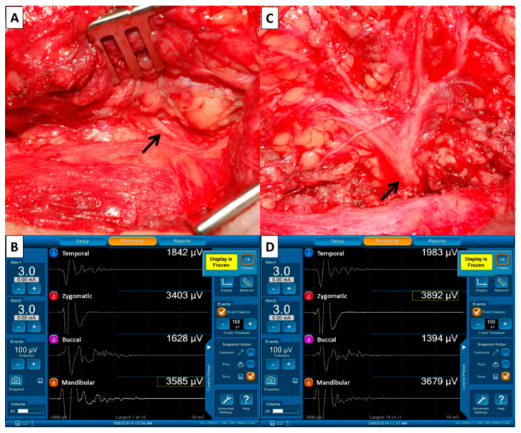
Figure 2.
Pre- and post-dissection facial nerve (FN) signals to evaluate FN function. (A) The FN trunk (↑) was first identified during parotid gland dissection and we performed supramaximal stimulation of the trunk using 3–5 mA. (B) The four elicited EMG signals representing the function of FN branches were displayed on the monitoring screen. The four EMG signals were defined as F1 signals and were used as basic reference data before dissection of the FN branches. (C) The stimulus current (3–5 mA) was applied to the FN trunk (↑) after dissecting the FN branches and resecting the parotid tumor. (D) The four elicited EMG signals were defined as F2 signals. The EMG amplitudes on each channel of F1 and F2 signals were compared.

