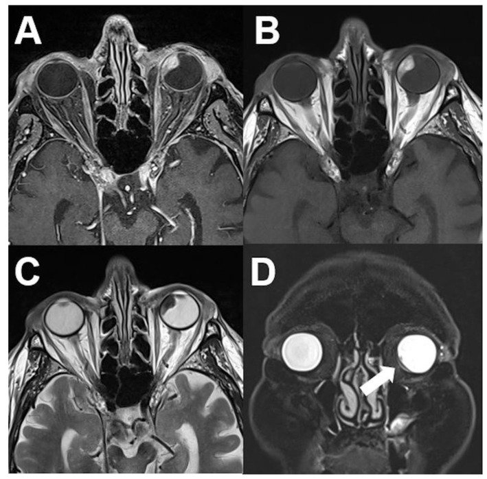
Figure 2.
Uveal melanoma. MRI of an 84-year-old male patient with uveal melanoma showing a well-defined tumor of the left nasal vitreous body. The tumor shows a hyperintense signal on unenhanced T1-weighted images (A), a diffuse enhancement on contrast-enhanced T1-weighted images (B), and a marked hypointense signal on T2-weighted images (C). Retinal detachment with subretinal fluid (arrows) is shown in the coronal view (fat saturated T2-weighted sequence) (D).

