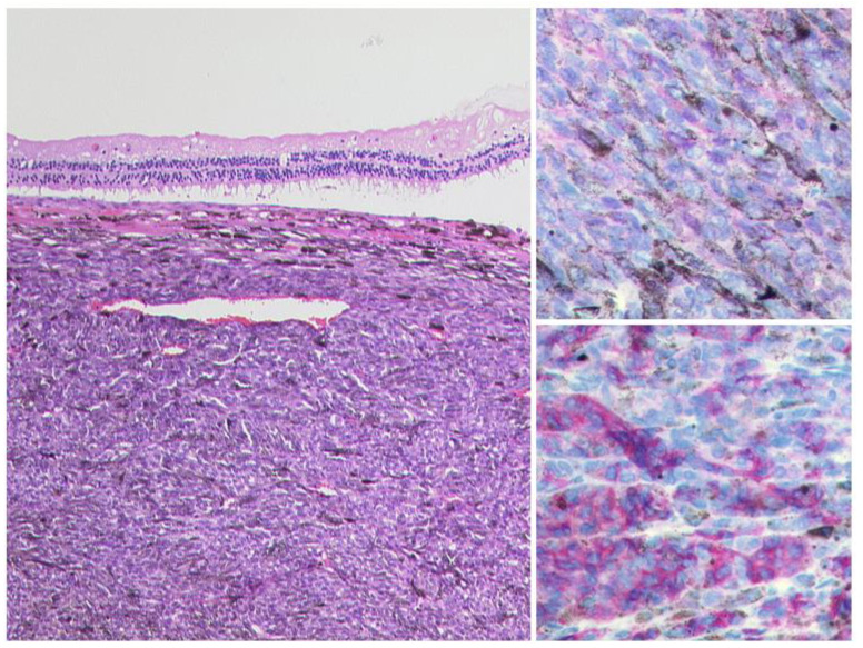
Figure 3.
Uveal Melanoma. Left: A pigmented tumour mass is seen beneath the artificially detached retina, HE, Original magnification 50:1. The tumour cells show a weak expression of S100 (upper right, original magnification 200:1) and a modest expression of Melan A (lower right, original magnification 50:1).

