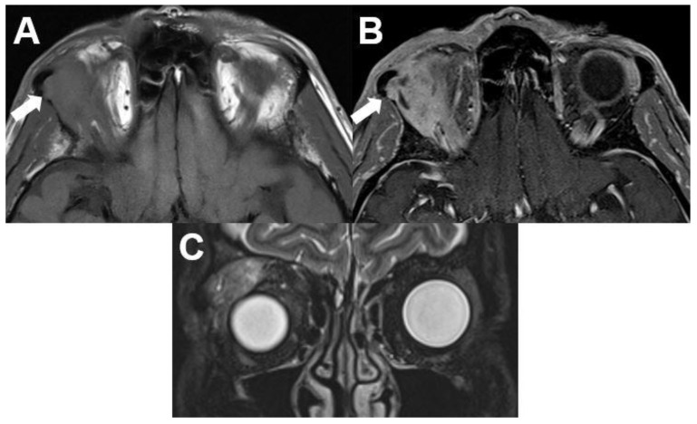
Figure 15.
Carcinoma of the lacrimal gland. MRI of a 59-year-old male patient with a biopsy-proven poorly differentiated carcinoma of the right lacrimal gland. T1-weighted axial images show an ill-defined mass of the right lateral cranial orbit with signal characteristics similar to surrounding muscles (A) and contrast enhancement (B). The adjacent zygomatic bone is infiltrated (arrow). On T2-weighted images of the coronal view, the tumor shows an inhomogeneous signal (C).

