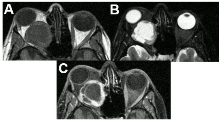
Figure 20.
Schwannoma of the optic nerve. MRI of a 36-year-old female presenting with a large retrobulbar tumor and exophthalmos. The tumor is hypointense on T1-weighted (A), hyperintense on T2-weighted sequences (B), and shows avid contrast enhancement of its solid parts (C). Note the cystic degeneration of the tumor. The tumor was surgically removed, and pathology revealed a schwannoma.

