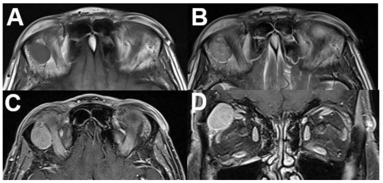
Figure 22.
Neurofibroma. MRI of a 59-year-old female patient with a tumor of the right-sided upper orbit. The tumor is located between the superior and lateral rectus muscle, showing T1-isointense (A) and slight T2-hyperintense (B) signal compared to the muscles. It shows heterogeneous contrast enhancement on axial (C) and coronal (D) views. In this case, diagnosis was proven after resection.

