FIG 2.
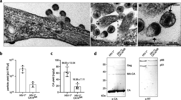
Production and characterization of click-labeled HIV-1 (HIV-1*CA14SiR). (a) Morphology of HIV-1*CA14ncAA assembly sites and particles. HEK293T cells were cotransfected with pNLC4-3*CA14TAG and pNESPylRS-eRF1dn-tRNA and grown in the presence of 500 μM CpK. At 44 h p.t., cells were fixed, embedded, and analyzed by thin-section EM as described in Materials and Methods. (b and c) Virus production. Click-labeled particles were prepared from the supernatant of HEK293T cells cotransfected with either pNLC4-3* or pNLC4-3*CA14TAG and pNESPylRS-eRF1dn-tRNA and grown in the presence of 500 μM CpK as described in Materials and Methods. Particle yield in the final preparations was determined via quantitation of RT activity (SG-PERT assay [79]) (b) and by determination of CA amounts using quantitative immunoblot as described in Materials and Methods (c). The graphs show mean values and SD from four independent experiments. (d) Immunoblot analysis of virus preparations. Five microliters of HIV-1* and HIV-1*CA14SiR particle lysates was separated by SDS-PAGE, and proteins were transferred to nitrocellulose membranes by semidry blotting. Viral proteins were detected using the indicated polyclonal antisera. Bound antibodies were detected by quantitative immunofluorescence with a Li-COR CLx infrared scanner, using secondary antibodies and protocols according to the manufacturer’s instructions. Positions corresponding to Gag/Gag-Pol and processing products are indicated at right.

