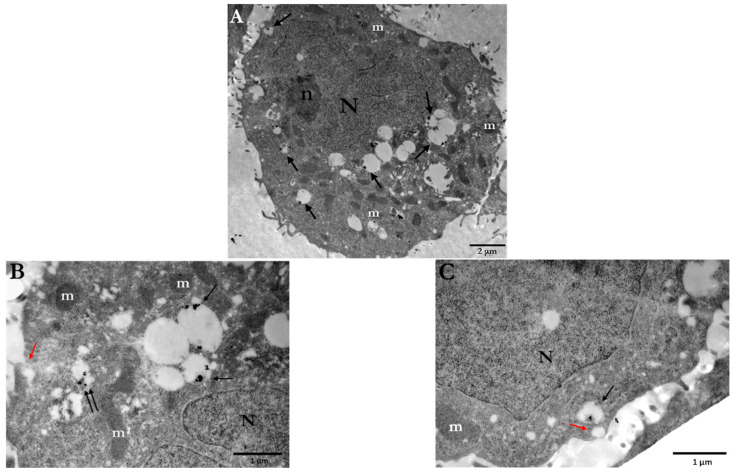
Figure 1.
Electron micrographs of PC3 cells indicating the cellular uptake of 15 nm Ct-AuNPs (10 μg/mL) after 24 h. (A) Arrows point to the different areas of the cytoplasm, where nanoparticles are located inside vesicles or autophagosomes. (B,C) Higher magnification of (A). Ct-AuNPs are located inside vesicles (single arrows) and autophagosomes (double arrows). Red arrows indicate vesicular formations of the plasma membrane by which NPs may be endocytosed or excreted by the cell. Scale bars: (A) 2 μm; (B) and (C) 1 μm. N: nucleus, n: nucleolus, m: mitochondrion.

