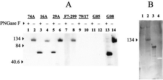
FIG. 3.
Western blot of anti-SU MAbs with nonreduced SU digested with PNGase F. (A) Affinity-purified SU incubated for 16 h at 37°C without PNGase F (lanes designated with minus signs) or with PNGase F (lanes designated with plus signs) was analyzed by Western blotting with anti-SU MAb or an irrelevant isotype control MAb under nonreducing conditions. Negative control serum from goat 8505 (G05) and positive control serum from goat 9808 (G08) were used at dilutions of 1:1,000. (B) SU incubated with or without PNGase F (lanes 1 and 2), positive control glycoprotein (transferrin) (lane 3), and unglysosylated negative control protein (creatinase) (lane 4) were subjected to SDS-PAGE and transferred to nitrocellulose. Glycan residues were oxidized with 10 mM NaIO4 in 0.1 M acetate buffer (pH 5.5) for 20 min at room temperature and stained by using a DIG glycan detection kit. Numbers to the left of each panel are in kilodaltons.

