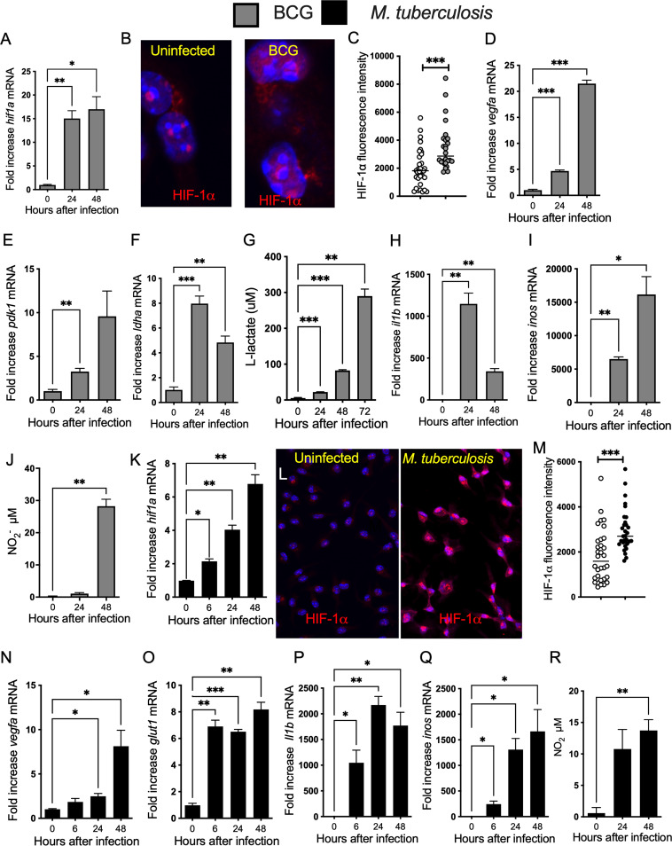
FIG 1.
HIF-1-regulated responses increase in mycobacteria-infected BMM. (A, D to F, H, I, K, and N to Q) Total RNA was extracted from triplicate independent cultures of BMM before and at different time points after infection with BCG (A, D to F, H, and I) or M. tuberculosis (K and N to Q) at an MOI of 5:1. The relative concentration of hif1a (A and K), vegfa (D and N), pdk1 (E), ldha (F), glut1 (O), il1b (H and P), and inos (I and Q) transcripts in relation to hprt mRNA levels in the same sample was determined by real-time PCR. Differences with noninfected controls are significant at *, P ≤ 0.05; **, P ≤ 0.01; and ***, P ≤ 0.001, Student's t test. (B, C, L, and M) Micrographs showing labeling for HIF-1α in BMM 24 h after either BCG (B) or M. tuberculosis (I) infection and in uninfected controls (DAPI was used for nuclear staining). The HIF-1α label intensity in BMM 24 h after infection with BCG (C) or M. tuberculosis (M) infected BMM was quantified using the Cell Profiler software. The quantification was performed in 3 independent samples (4 field of view per sample). The individual and the mean HIF-1α levels per cell in one field of view are shown. Differences are significant at ***, P ≤ 0.001, unpaired Student's t test. (G) The lactate concentration accumulated in the supernatants from BMM infected with M. bovis BCG (MOI 1:1) was measured by lactate dehydrogenase assay. The data represent the results of triplicate independent cultures ± SEM. A representative of 2 experiments is shown. Differences with noninfected controls are significant at **, P ≤ 0.01 and ***, P ≤ 0.001, unpaired Student's t test. (I and R) The concentration of nitrite in the supernatants of either M. tuberculosis (I) or BCG-infected (R) BMM was measured at different times after infection. The mean levels of NO2− ± SEM in independent triplicates were measured by Griess assay.

