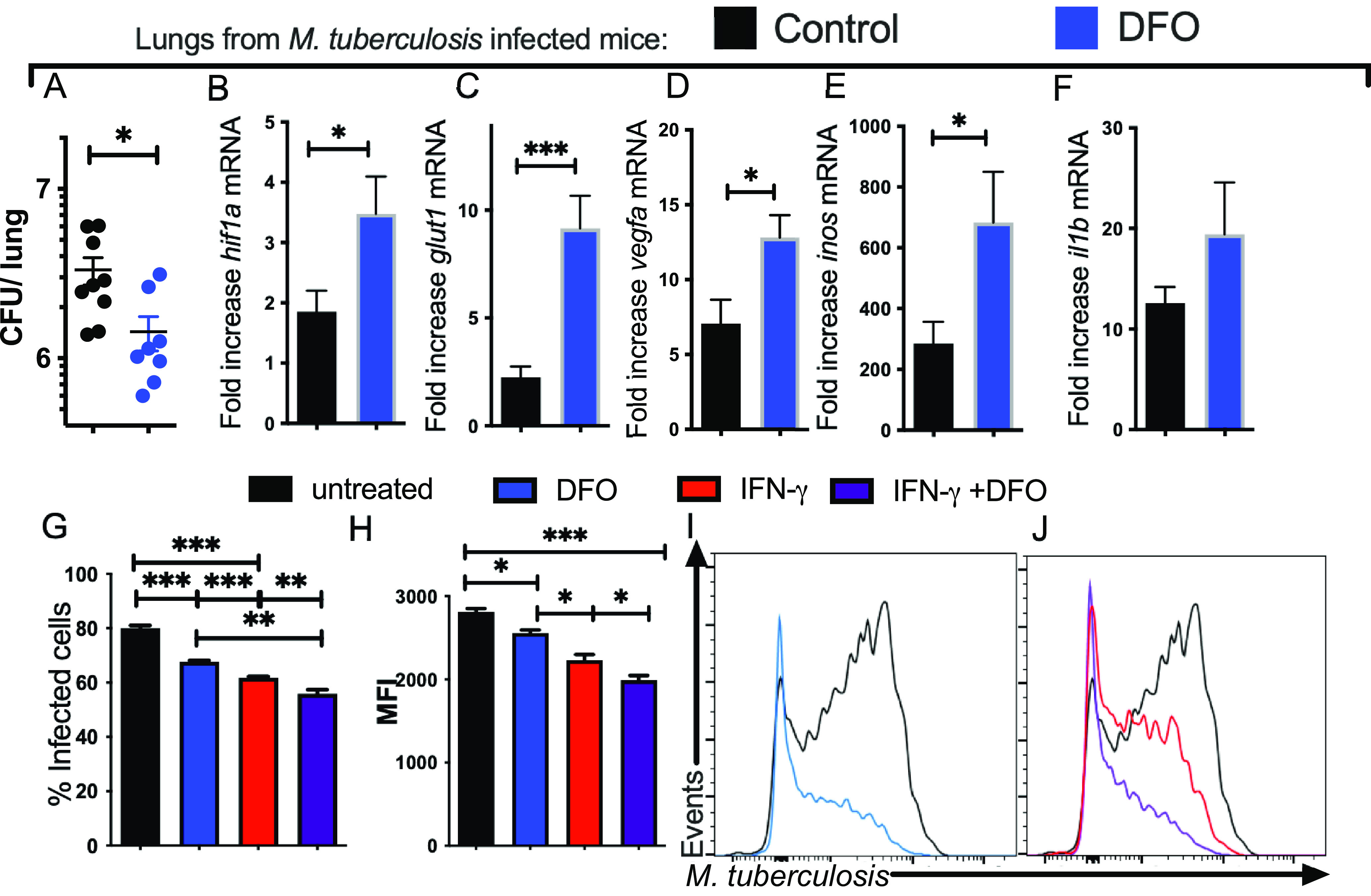
FIG 3.

DFO treatment of BMM improves the intracellular growth control of M. Tuberculosis. (A) C57BL/6 mice were treated with 400 mg/kg DFO i.p. every other day after aerosol infection with M. tuberculosis for 12 weeks. The group median and the individual CFU per lung of DFO-treated and control mice (n = 9 per group) are depicted (median CFU/lung: control 2.80·106; DFO treated: 7·106). Differences in CFU between DFO-treated and untreated groups are significant (*, P < 0.05, Mann-Whitney U test). (B to F) The mean relative levels ± SEM of hif1a (B), glut1 (C), vegfa (D), inos (E), and il1b (F) transcripts in lungs of DFO-treated or control mice (n = 5 per group) were measured by real-time PCR. The results were normalized with the level of a transcript in the lung of an untreated and uninfected control animal. Differences with the untreated group are significant at *, P ≤ 0.05 and ***, P ≤ 0.001, Student's t test with Welch’s correction. (G to J) A group of BMM was treated with IFN-γ starting 48 h before M. tuberculosis-gfp infection (MOI 1:1). Groups of IFN-γ-activated and control BMM were incubated with DFO 4 h after infection or left untreated. The mean percentage of infected BMM (G) and the mean M. tuberculosis-gfp MFI gated on infected cells (H) determined 5 days after infection and representative histograms are shown (I and J). Differences are significant at *, P ≤ 0.05; **, P ≤ 0.01; and ***, P ≤ 0.001, one-way ANOVA test with Welch’s correction.
