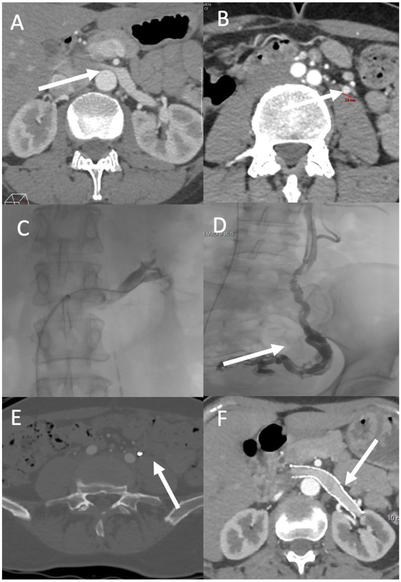
Figure 3.
PVD affecting zones 1 and 2. A 49-year-old-woman with left flank pain (VAS pain score = 7), hematuria, and left chronic pelvic pain exacerbated by prolonged standing (VAS pain score = 5). (A) CT shows compression of the left renal vein over the abdominal aorta (arrow) and (B) a dilated left ovarian vein (diameter = 8 mm; arrow). Venography demonstrates attenuation over the abdominal aorta with a venous pressure gradient of 6 between the vena cava and left renal vein (C), and an incompetent left ovarian vein feeding a pelvic varicose vein (arrow) (D). Three months after treatment, CT shows successful embolization (using Onyx) of the left ovarian vein ((E), arrow) and patency of the left ovarian vein stent ((F), arrow). The left flank and chronic pelvic VAS pain scores decreased to 3 and 1, respectively.

