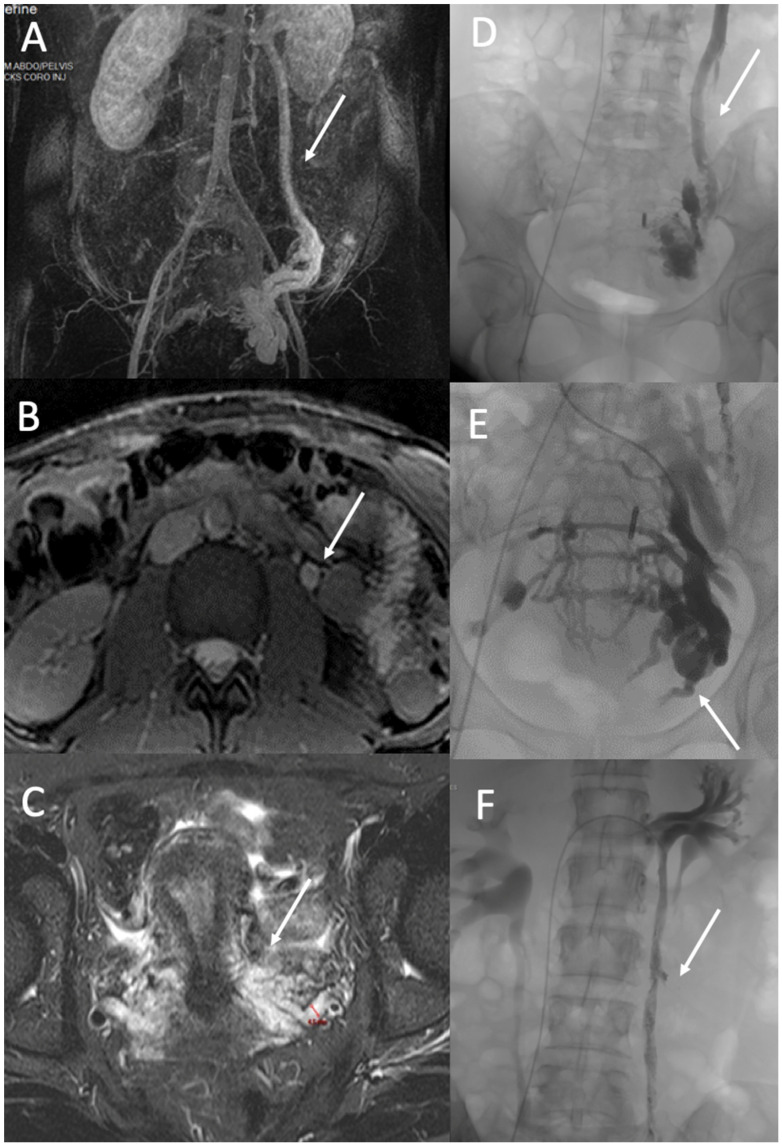
Figure 4.
PVD of Zone 2. A 37-year-old woman with a typical pelvic congestion syndrome. (A). MR angiography image in the coronal plane shows incompetent and dilated left ovarian vein (arrow) and left pelvic veins with right periuterine varicose vein (B). True fast imaging with steady-state-free precession (TRUFI) demonstrated a dilated left ovarian vein (arrow) (10 mm) (C), and T2 STIR MR images (C) in an axial plane demonstrated dilated pelvic veins up to 8 mm (arrow). Phlebography shows incompetent left ovarian vein (arrow) (D) and incompetent internal iliac veins with periuterine and uterine varicose (arrow) (E). Treatment consisted of embolization with Onyx and Aetotoxisclerol of ovarian vein (arrow) (F) and pelvic varicosities.

