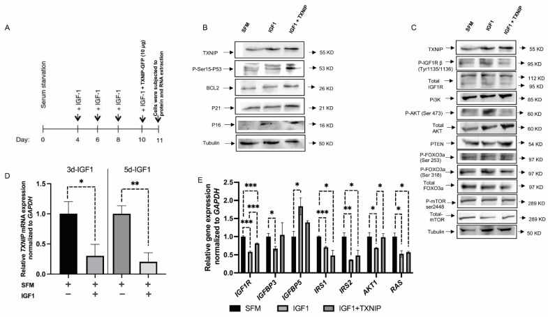
Figure 4.
Effect of TXNIP on prolonged IGF1-induced senescence. (A) Schematic representation of senescence induction by prolonged IGF1 treatment. (B,C) Primary skin fibroblasts were treated with IGF1 for 11 days, transfected with a TXNIP-GFP vector during the last 24 h of IGF1 treatment, or serum-starved for the entire period and transfected with an empty pcDNA-GFP plasmid (SFM). At the end of the incubation period, cells were harvested and the indicated proteins were measured by Western blots. (D) Relative TXNIP mRNA levels upon 3 or 5 days of IGF1 treatment with IGF1 (or serum starved controls). (E) Gene expression analysis in IGF1-induced premature senescence with or without TXNIP-GFP transfection. GAPDH mRNA levels served as an internal control. * p-value ≤ 0.05; ** p-value ≤ 0.01; *** p-value ≤ 0.001.

