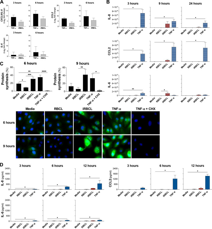
FIG 6.
TNF-α and P. falciparum-iRBC lysates induce transcriptionally similar, but translationally different levels of cytokines and chemokines. (A to D) HBMEC monolayers were incubated with media, TNF-α (1 ng/mL), RBCLs (8 × 10⁶/cm2), or P. falciparum-iRBCLs (8 × 10⁶/cm2) for the indicated times. (A) Quantitative PCR for each time point was conducted to determine the expression levels of IL8, CCL2, and IL6. The fold change of TNF-α relative to medium (black bars) and iRBCLs relative to RBCL (gray bars) is shown. (B) Lysates of HBMECs after the incubation were used to determine intracellular protein levels of IL-8, CCL2, and IL-6. Cytokine and chemokine levels were normalized to the total protein level of the cell lysate. (C) Changes in protein synthesis were determined by the incorporation of Click-iT HPG-Alexa Fluor 488 measured by flow cytometry and visualized by immunofluorescence microscopy. Representative images are shown. HPG is in green, and nuclei are in blue. TNF-α with cycloheximide (CHX) (100 μg/mL) was included as a control. (D) Medium of HBMEC cultures was collected to determine secreted protein levels of IL-8, CCL2, and IL-6. Results are the average from at least 3 independent experiments with standard deviation. Statistical significance was determined by (C) one-way ANOVA with Tukey’s multiple-comparison test or (A, B, and D) Kruskal-Wallis test with Dunn’s multiple-comparison test (*, P < 0.05; **, P < 0.01; ***, P < 0.001; ****, P < 0.0001).

