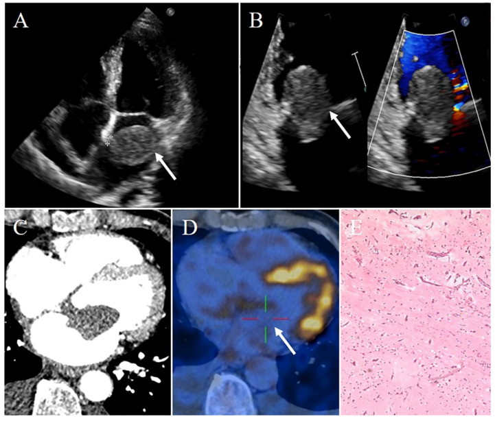
Figure 1.
Left atrial myxoma in a 69-year-old man presenting with chest tightness and shortness of breath. (A) Transthoracic echocardiography showing a pedunculated mobile heterogeneous echogenic mass attached to the interatrial septum. This mass locates in the LA. (B) Part of this mass protruding into the LV through the mitral valve orifice in diastole, leading to stenosis of the mitral valve orifice. (C) Contrast-enhanced CT demonstrating a relative low density well-circumscribed mass originating from the interatrial septum, with absent of enhancement. (D) PET-CT imaging revealing the radionuclides slightly concentrated in the mass (SUVmax 4.6). (E) Pathology confirming myxoma. White arrows pointing to the left atrial myxoma and † marking stalk.

