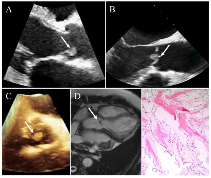
Figure 2.
Incidental finding of a papillary fibroelastoma in a 62-year-old man. (A) A small mobile solid mass attaching to the aortic valve on transthoracic echocardiography. (B) Transesophageal echocardiography clearly showing the mass of aortic valve. (C) Three-dimensional echocardiography clearly visualizing the location and size of the mass. (D) CMR demonstrating a round, small, homogeneous mass attached to aortic valvular leaflet. (E) Pathology confirming papillary fibroelastoma. White arrows representing the papillary fibroelastoma.

