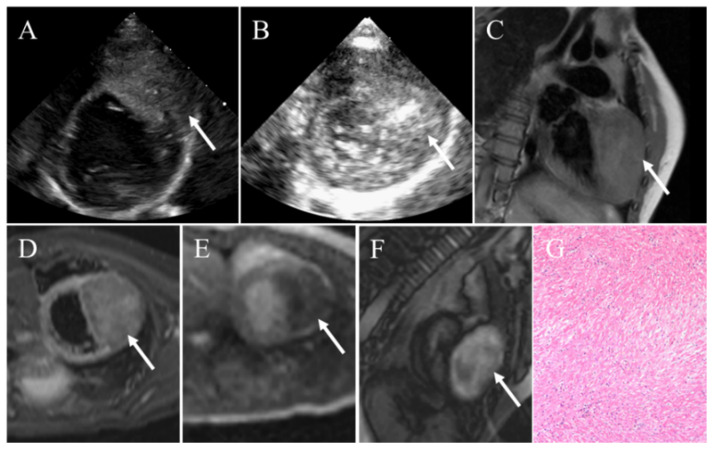
Figure 4.
Cardiac fibroma in a 3-year-old child manifesting as palpitation and cough. (A) Transthoracic echocardiography demonstrating a large heterogeneous intramyocardial mass with sporadic calcific. (B) Contrast echocardiography revealing slight enhancement of contrast agent within the mass. (C) CMR showing an intramyocardial mass presenting iso-intense on T1-weighted images. (D) On T2-weighted images, the mass appearing slight hyper-intense. (E) The mass presenting as hypoperfusion on resting first-pass perfusion images. (F) LGE imaging revealing the mass appeared as obviously inhomogeneous high signal intensity relative to the myocardium. (G) Pathology confirming fibroma. White arrows pointing to the cardiac fibroma.

