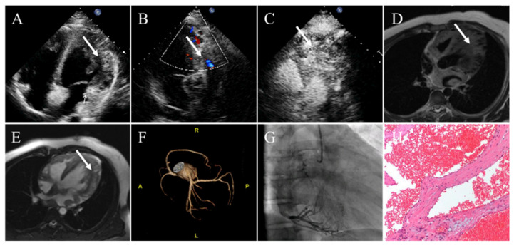
Figure 5.
Cardiac cavernous hemangioma in a 16-year-old man presenting with palpitation. (A) Transthoracic echocardiography showing a heterogenous echogenic mass in the lateral wall of the LV. (B) Color Doppler flow imaging revealing coronary artery blood flow within the mass. (C) Contrast echocardiography demonstrating enhancement of contrast agent within the mass. (D) The left ventricular wall appearing inhomogeneous thickening with local nodules and diffuse edema on T2-weighted images. (E) Late gadolinium enhancement imaging demonstrating that the lateral wall of the left ventricle presented as obviously inhomogeneous hyperintense. (F) On contrast-enhanced CT, the coronary artery branches increased in the left ventricular myocardium. (G) Coronary angiography confirms the coronary arteries give off many branches and myocardial obviously staining in arterial phase. (H) Pathology confirming cavernous hemangioma. White arrows representing the cardiac cavernous hemangioma.

