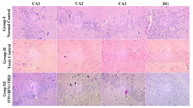
Figure 5.
The H/E-stained hippocampal section visualized under 40× shows normal arrangements and distributions in the normal control group (Group-I). Plenty of neuronal loss, degeneration and death from electroshock observed in Group-II. Slight cellular changes in CA1, CA2, CA3, DG of the SVP (150 mg/kg) + BFN (5 mg/kg) + THQ (40 mg/kg) injected rats are indicative of minimal neuronal degeneration and nuclear pyknosis in Group-XII.

