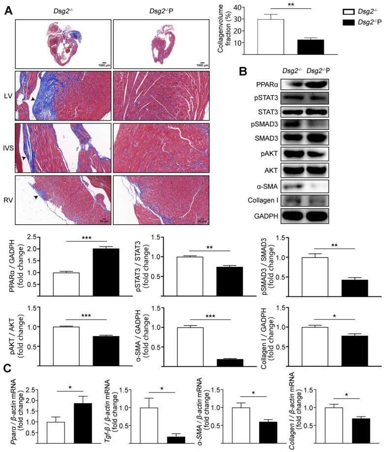
Figure 4.
AAV9-Pparα alleviated cardiac fibrosis in CS-Dsg2−/− mice. (A) Masson staining of heart sections in CS-Dsg2−/− mice and CS-Dsg2−/− mice received AAV9-Pparα (Dsg2−/−P). Collagen volume fraction in the hearts of CS-Dsg2−/− and Dsg2−/−P mice were assessed. (B) Representative Western blots from ventricles of CS-Dsg2−/− and Dsg2−/−P mice. PPARα, pSTAT3, pSMAD3, pAKT, α-SMA, and Collagen I were detected using specific antibodies. STAT3, SMAD3, AKT, and GAPDH were used as loading controls. (C) Results of quantitative PCR analysis of PPARα, TGF-β, α-SMA, and Collagen I mRNA levels in mouse ventricles are expressed as fold change of control using β-actin as loading control. Results are expressed as mean values ± SEM. n = 6. * p < 0.05, ** p < 0.01, *** p < 0.001 vs. CS-Dsg2−/−.

