FIG 2.

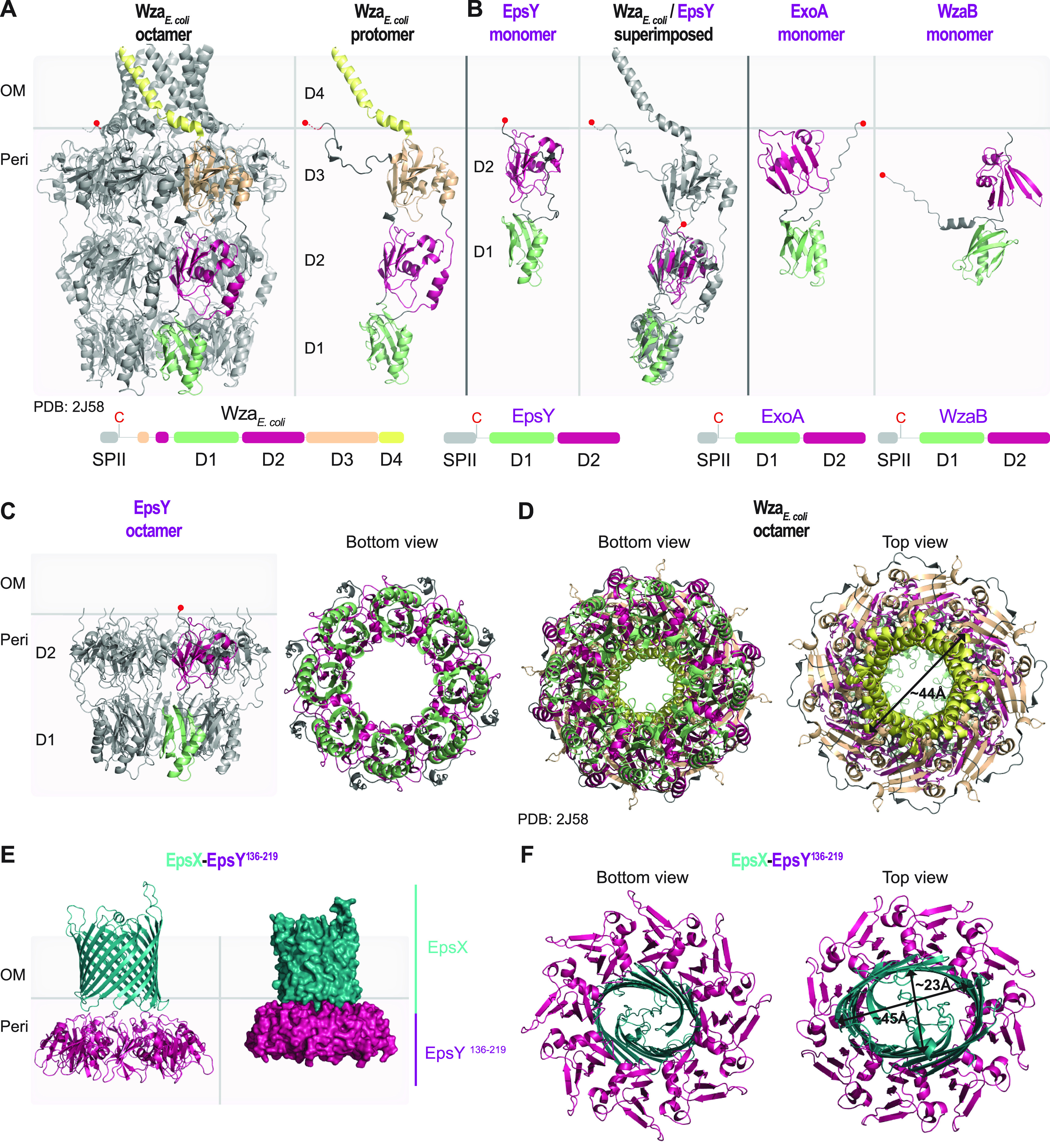
Structural characterization of the EpsY D1D2OPX protein alone and in complex with EpsX, its partner 18-stranded β-barrel protein. (A) Structure of WzaE. coli. Left panel, the solved structure of octameric Wza (PDB 2J58) (7). Right panel, an individual Wza protomer. The four domains of Wza are labeled D1 to D4. Light green, D1; dark pink, D2; light orange, D3; yellow, D4. The acylated N-terminal cysteine is indicated by a red circle and placed at the inner leaflet of the OM. Lower panel, domain organization of Wza. (B) AlphaFold model of EpsY. Left panel, lateral view of EpsY monomer as predicted by AlphaFold. Right panel, superimposition of a Wza protomer from the solved structure (gray) and the EpsY model. The EpsY monomer aligns to the Wza protomer with an RMSD of 3.306 Å over 904 Cα. Right panels, AlphaFold models of ExoA and WzaB monomers. In all three AlphaFold models, the two domains are labeled D1 and D2 and colored according to the homologous domains in Wza. The acylated N-terminal cysteine is indicated by a red circle; note that the acylated N-terminal cysteine of WzaB is not modeled “on top” of D2, but the confidence in the relative position of this residue is low (Fig. S3C). Model rank 1 is shown for all structures. Lower panels, domain organization of EpsY, ExoA, and WzaB. SPII, type 2 signal peptide. (C) AlphaFold-Multimer model of octameric EpsY. Left panel, one protomer is colored as described in the legend for panel B. Right panel, bottom view of octameric EpsY with all protomers colored as described for panel B. Model rank 1 is shown. (D) Structure of Wza. All eight protomers are colored as described in the legend for panel A. An arrow indicates the external diameter of the α-helical pore. In the bottom view, the tyrosine residues that form the so-called tyrosine ring are present in the loops extending into the central channel. (E and F) AlphaFold-Multimer model of a heterocomplex of octameric EpsY136-219 and an EpsX monomer. In the heterocomplex, the EpsY136-219 octamer is colored as D2 as described for panel B, and EpsX is colored teal. (E) The right panel is a surface-rendered representation. Model rank 1 is shown. (F) Arrows indicate the diameter of the β-barrel. Model rank 1 is shown.
