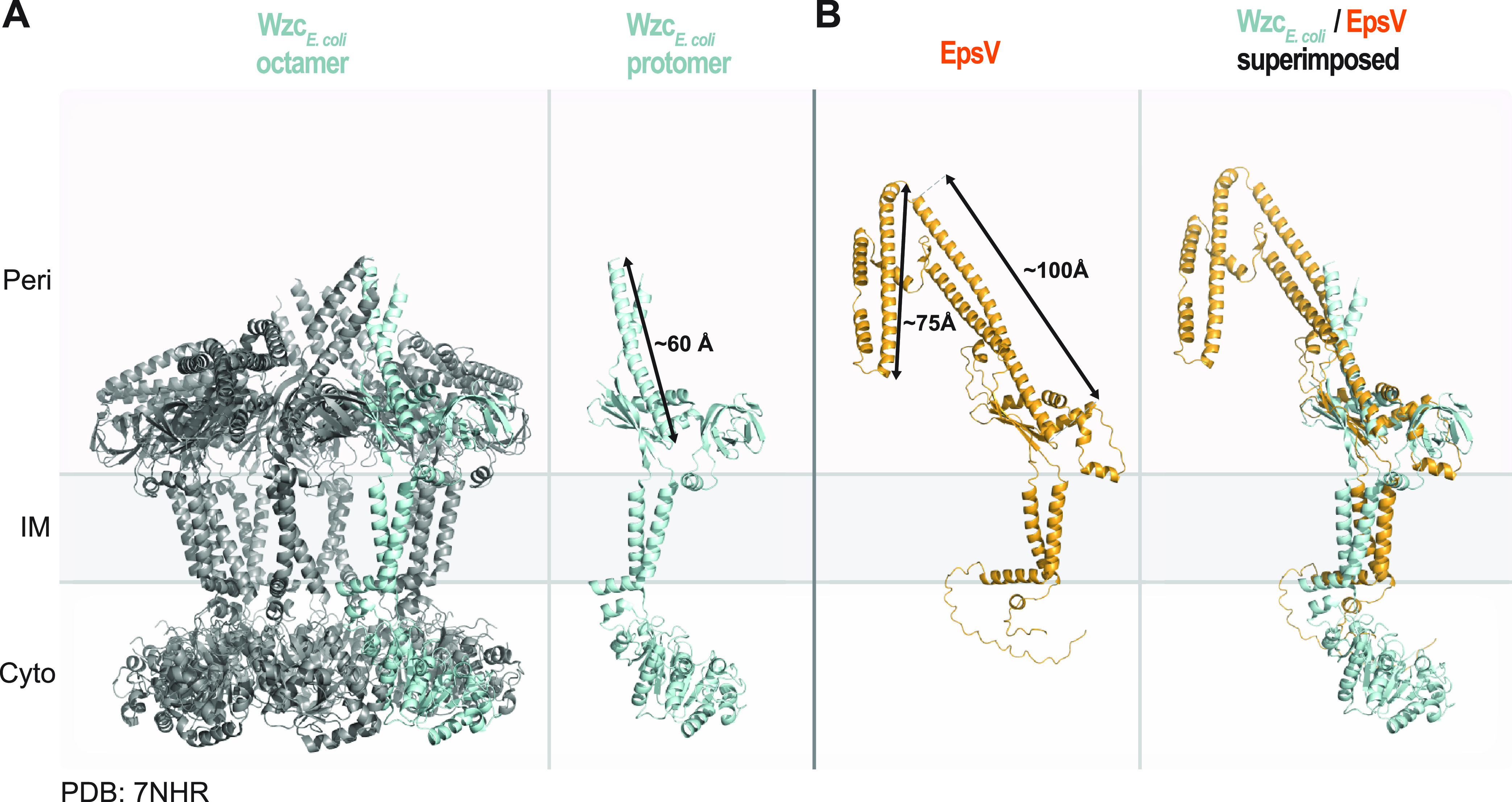
FIG 3.

Structural characterization of the PCP EpsV. (A) Solved structure of octameric WzcE. coli (PDB 7NHR) (30). Left panel, the protein is colored in gray, and one protomer is colored in light blue. Right panel, individual Wzc protomer in class 2 conformation (30). Note that individual protomers have different conformations in the octamer. An arrow indicates the length of the extended α-helical stretch. (B) AlphaFold model of EpsV. Arrows indicate the lengths of the α-helical stretches. Model rank 1 is shown. Right panel, superimposition of a protomer from the solved structure of Wzc and the EpsV model. EpsV aligns to Wzc with an RMSD of 4.610 Å over 727 Cα.
