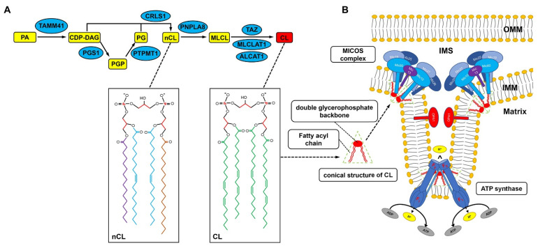
Figure 1.
Schematic diagram depicting the synthesis, remodeling, and localization of CL inside the mitochondria. (A) Inside the mitochondria of mammalian cells, the biosynthesis of CL begins with the conversion of phosphatidic acid (PA) to CDP-DAG catalyzed by TAMM41, followed by the transfer of a phosphatidyl group from CDP-DAG to glycerol-3-phosphate to form PGP, a process catalyzed by PGS1. PGP is subsequently dephosphorylated by PTPMT1 to generate PG, and CL is synthesized by CRLS1 through an irreversible condensation reaction of PG and CDP-DAG. The newly synthesized CL (nCL) contains a mixture of fatty acyl chains differing in length and saturation. To become mature CL, nCL undergoes a series of deacylations and reacylations, a process called remodeling. nCL is first deacylated to monolysocardiolipin (MLCL) by phospholipase A2 PNPLA8 and then reacylated by acyltransferases, including tafazzin (TAZ), acyl-CoA: lysocardiolipin acyltransferase 1 (ALCAT1), and MLCL acyltransferase-1 (MLCL AT1) to achieve a final symmetric acyl composition in mature CL. (B) The tetra-acyl chains and the negatively charged polar moiety give CL a conical shape, which results in CL causing negative curvature in the mitochondrial membrane and the IMM bending, forming the cristae. In addition, the mitochondrial contact site and cristae-organizing system (MICOS) complex and ATP synthase are also essential for the formation of mitochondrial cristae.

