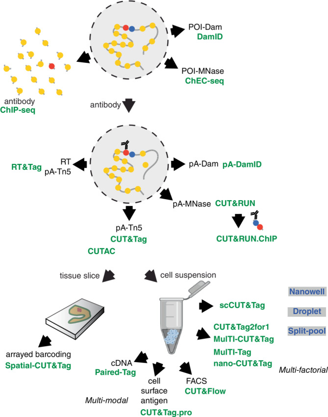
FIGURE 2.

Schematic of the in situ chromatin profiling methods described in this survey. Methods are indicated in green, with their characteristic reagents or features in black. POI, protein of interest. Chromatin features within a permeabilized nucleus (dashed circle) are indicated in yellow, red, and blue. Platforms that have been used for single‐cell scCUT&Tag and multifactorial methods are shown in gray boxes.
