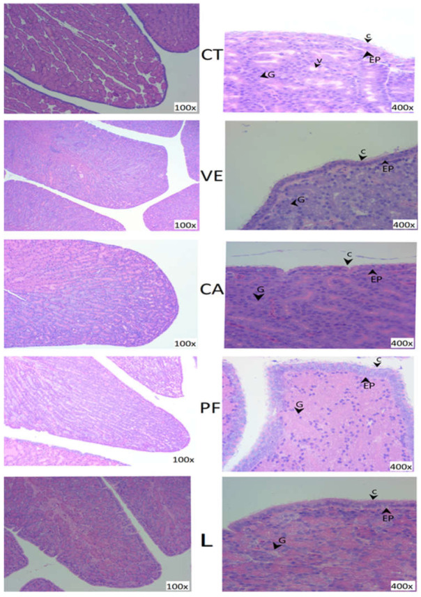
Figure 2.
Hematoxylin-and-eosin-stained images of the cross section (100× magnification) or longitudinal section (400× magnification) of the magnum in laying hens (36 wk) after a 10-wk treatment period. EP = epithelium line of the mucosa; C = cilia; G = tubular glands; V = vacuole; control = CT; VE 0.02% = vitamin E; CA 0.24% = chlorogenic acid; PF 0.05% = polyphenol; and L 0.03% = lutein.

