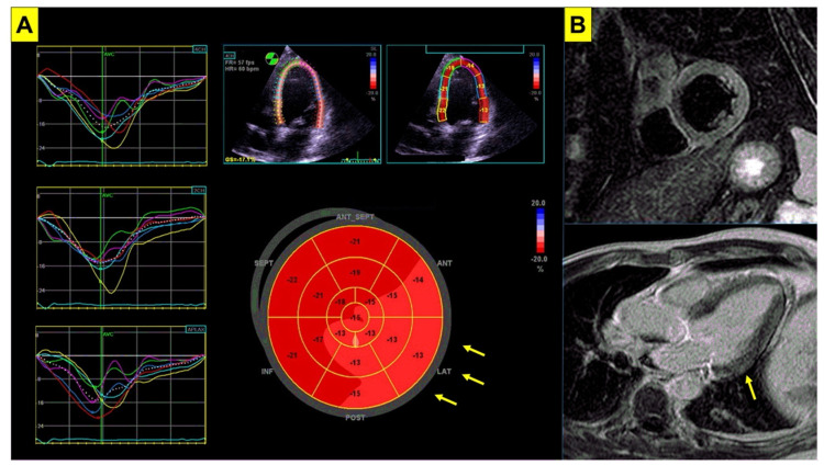
Figure 2.
Imaging abnormalities in Brugada syndrome. Subtle imaging abnormalities associated with BrS are shown. Panel (A) echocardiogram of a patient with genetically proven BrS. Despite normal left ventricular systolic function (LVEF = 62%), impairment in global longitudinal strain is shown (GLS = −16%, nv < −20%) mainly involving the lateral wall (arrows). Panel (B) cardiac magnetic resonance in the same patient shows slight hyperintensity in T2-weighted short tau inversion recovery sequences (STIR, upper panel) involving the inferolateral basal segment of the left ventricular wall, and focal late gadolinium enhancement (LGE, lower panel) involving the basal segment of the lateral wall (arrow). BrS = Brugada syndrome; LVEF = left ventricular ejection fraction.

