FIG 6.
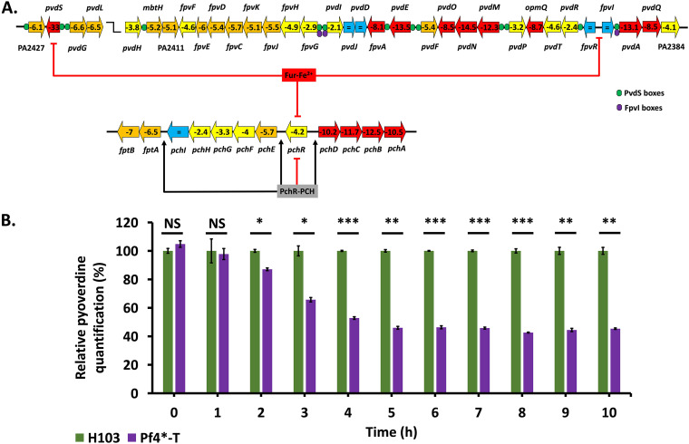
Pyoverdine production upon Pf4* infection. (A) Pyoverdine-encoding and pyochelin-encoding gene organization and regulation of the operons. The expression of each gene upon Pf4* infection is indicated inside arrows, and the colors indicate the following: red, >10-fold underexpressed; orange, downregulation between 5- and 10-fold; yellow, underexpression between 2- and 5-fold; and blue, not differentially expressed. PvdS binding sites are indicated by green circles, and FpvI binding sites are indicated by violet circles. The pvdS, fpvR, and fpvI genes are repressed by Fur-Fe2+, as well as the pchR gene. The product PchR, once bound to pyochelin (PCH), represses its own expression, whereas it activates the fptAB, pchEFGHI, and pchDCBA operons. See the text for more details. (B) Relative pyoverdine quantification (± the SEM) of the H103 (in green) and Pf4*-T (in purple) conditions in iron-poor medium (CAA). Pyoverdine quantifications were normalized with A580. Pyoverdine quantification was assayed three times independently. Statistics were determined using a paired (two-sample) two-tailed t test (NS, P > 0.05; *, P < 0.05; **, P < 0.01; ***, P < 0.001).

