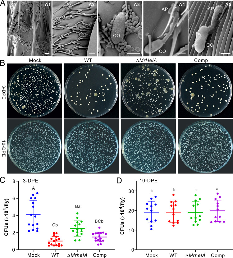
FIG 4.
Manipulation of Drosophila cuticular bacterial loads by different fungal strains. (A) SEM observation of Drosophila surfaces. Dense loads of bacterial cells were observed on the tarsal segments (A1) and abdomen (A2) of 10-DPE flies. Once in contact with multiple bacterial spores, germination of the conidium (CO) was inhibited (A3). Otherwise, spores could germinate to form appressoria (AP) with no bacterial contact (A4) or in contact with a few bacterial cells (A5). Bar, 5 μm. (B) CFU formation of the bacteria washed from the body surfaces of 3-DPE (top) and 10-DPE (bottom) male flies after treatment with different fungal strains. The wash solutions (10 flies in 1 mL of PBS buffer) were diluted 10 times prior to being plated on the LB agars. (C and D) Determination of the fly surface bacterial CFU after topical infection of 3-DPE (C) and 10-DPE (D) male flies. After treatment for 16 h, the flies were anesthetized and washed for plating and CFU counting. Ten male flies were collected and washed as an independent replicate. Values are means and SD. One-way ANOVA was conducted to determine the difference between treatments. Different letters above values show the difference with P values of <0.01 (capital) and <0.05 (lowercase).

