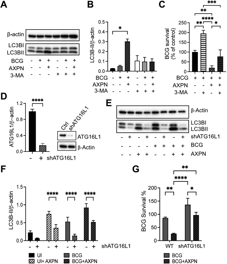
FIG 3.
Autophagy contributes to Amoxapine-mediated suppression of intracellular survival of BCG in macrophages. (A) RAW 264.7 cells were infected with BCG for 3 h and treated with 10 μM Amoxapine for 24 h in the presence or absence of 5 mM 3-MA. Western blots detected LC3B-II and actin levels. (B) Image J was used to quantify protein expression levels. (C) Intracellular BCG survival was enumerated by CFU in RAW 264.7 with BCG infection and 10 μM Amoxapine treatment in the presence or absence of 3-MA at day 2 postinfection. (D) ATG16L1 expression levels were detected by Western blots in control and shATG16L1 knockdown of RAW264.7 cells. (E to F) Western blots and quantification of LC3B-II levels in control and shATG16L1 knockdown of RAW264.7 cells with BCG infection and treatment with 10 μM Amoxapine at 24 h postinfection. (G) Intracellular survival of BCG was enumerated by CFU in control and shATG16L1 knockdown of RAW264.7 cells with BCG infection and 10 μM Amoxapine treatment at day 1 postinfection. Data represent means ± standard deviations for three independent experiments. One-way ANOVA with Dunnett’s multiple-comparison test was used for statistical analysis to compare the 3-MA-treated group to the untreated group or ATG16L1 knockdown groups to the control group. *, P < 0.05; **, P < 0.01; ****, P < 0.0001.

