Figure 1.
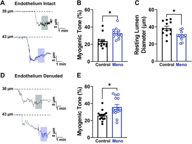
Menopause increases myogenic tone and decreases resting lumen diameter in C57BL/6 mice. A: representative traces of the lumen diameter of parenchymal arterioles from control (top, black) and menopause (Meno) (bottom, blue) mice showing increased myogenic tone, observed as a larger contraction to the physiological intraluminal pressure of 40 mmHg. Shaded areas indicate where measurements were taken. B: summary data showing that Meno induced a statistically significant increase in spontaneous myogenic tone of isolated, pressurized parenchymal arterioles. C: consequently, the resting lumen diameter of parenchymal arterioles isolated from Meno mice was significantly reduced compared with those isolated from control mice. Data are means ± SE; N = 12 arterioles from 8 different mice. *P < 0.05, 2-tailed Student’s t test. D and E: removal of the endothelium did not affect the increased myogenic tone observed in Meno mice, as shown by the representative traces (D) and the summary bar graphs (E). Data are means ± SE; N = 14 arterioles from 7 different mice. *P < 0.05, 2-tailed Student’s t test.

