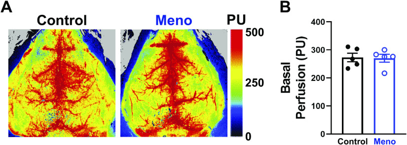
Figure 6.
Basal cerebral perfusion is unaltered by menopause. A: representative heat maps from laser-speckle contrast imaging showing no change in basal cerebral perfusion between control (left) and menopause (Meno; right) mice. PU, perfusion units. B: summary bar graph showing that basal cerebral perfusion is not significantly altered by menopause in mice. Data are means ± SE; N = 5 mice/group, analyzed by 2-tailed Student’s t test.

