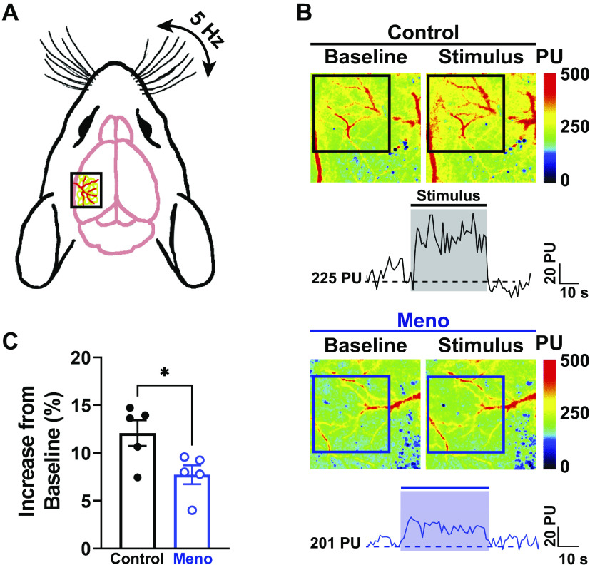
Figure 7.
Menopause impairs neurovascular coupling. A: diagram of a mouse head showing laser-speckle contrast imaging recording on top of the whisker-barrel cortex (black box) during contralateral whisker stimulation. B: representative heat maps and traces at baseline (left) and during mechanical stimulation of the whiskers (right). Squares show the area where measurements were performed (whisker-barrel cortex). Note the increase in perfusion (warmer colors) in the microcirculation of control females (top), which was blunted in menopause (Meno) (bottom). This can also be observed in the representative traces shown below the images. The shaded areas on the traces show the region where the perfusion measurements were taken. PU, perfusion units. C: summary data showing a significantly blunted hemodynamic response upon whisker stimulation in Meno. Data are means ± SE; N = 5 mice/group. *P < 0.05, 2-tailed Student’s t test.

