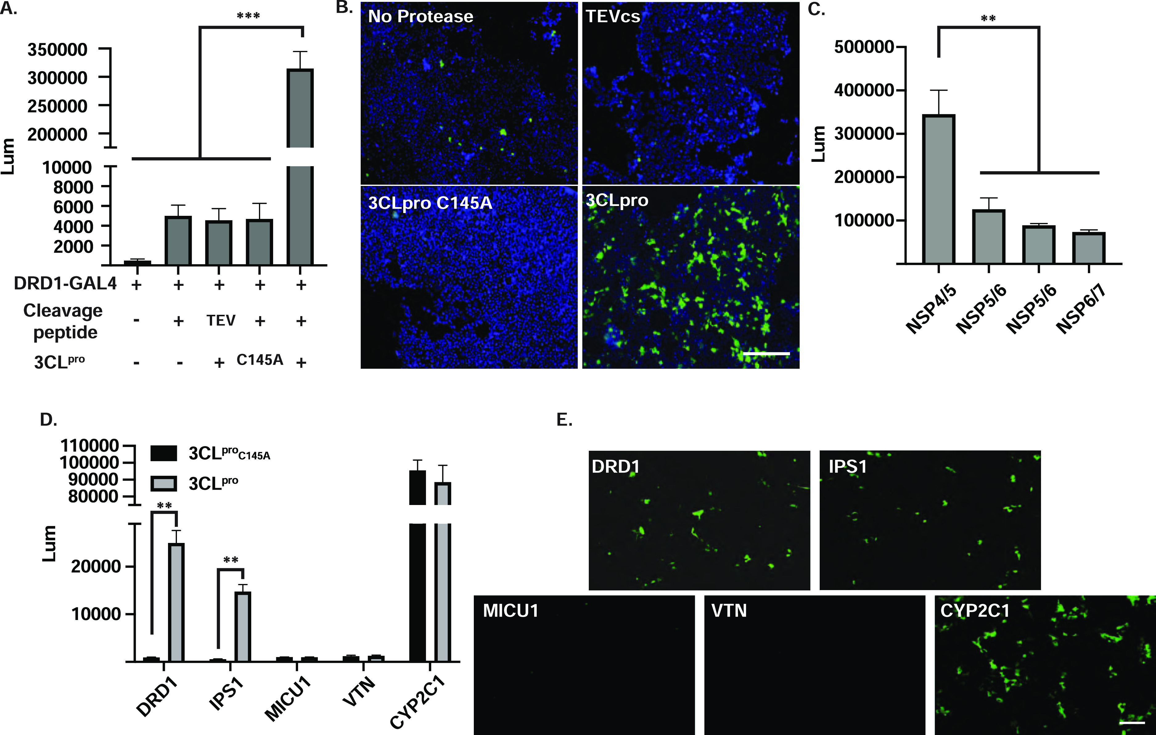
Figure 2.

Validation of the SARS-CoV-2 3CLpro live-cell reporter. (A) HEK293T cells were transfected with reporter components as stated in the panel, and luciferase activity was quantified 48 h after transfection. Significant increase was detected only in cells that expressed catalytically active 3CLpro together with the corresponding cleavage site. Data was plotted as mean ± SD. Statistical analysis was performed using a two-tailed Student’s t-test, N = 3, *** = p ≤ 0.005. (B) Representative fluorescence images of citrine expression in HEK293T cells 48 h after transfection. Scale bar, 100 μm. (C) HEK293T cells were transfected with GAL4-DRD1 fusion with different 3CLpro linkers corresponding to the viral NSP’s junction sites as stated in the panel. Luciferase activity was quantified 48 h after transfection. N = 3, data was plotted as mean ± SD. (D) 3CLpro cleavage efficiency was evaluated with different GAL4 fusion proteins as stated in the graph (NSP4/5 linker was used in all the constructs) and compared to the catalytically inactive 3CLproC145A. N = 3, data was plotted as mean ± SD. (E) Representative fluorescence images of citrine expression in HEK293T cells 48 h after transfection 3CLpro and different GAL4 fusion proteins. Scale bar, 100 μm.
