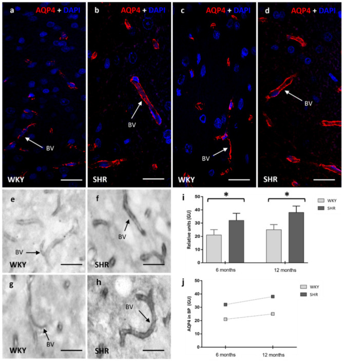
Figure 3.

Confocal and optical microscopy images of AQP4 in the blood vessels of brain parenchyma of WKY and SHR rats. A significant increase of AQP4 was observed in SHR in the 6 months group when compared to WKY (a,b,e,f), in the 12 months group was found a more significant presence of AQP4 in the SHR (c,d,g,h) rats. Stain intensities in relative units for AQP4 (i,j) are represented as the means ± SD (n = five animals per group). The differences between WKY and SHR were significant when applied to a One-way ANOVA test with post hoc analysis using the Tukey post hoc test (* p < 0.05). BV: blood vessel; GU: grey units. Scale bars 10 μm.
