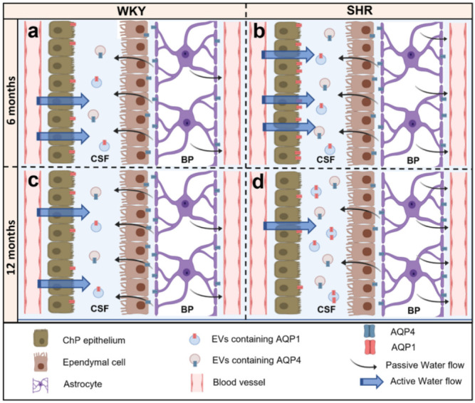
Figure 5.

Schematic representation of the expression of AQP1 and AQP4 in 6 months and 12 months WKY and SHR rats. In 6 months and 12 months WKY (a,c), rat expression of AQP 1 is detected in the apical pole of the ChP epithelium associated with active production of CSF. It is also found in the CSF, most likely associated with EVs. Per contra, AQP4 is expressed in the astrocyte end-feet and the basolateral membrane of the ependymal cells associated with brain CSF homeostasis. In addition, it is found in the CSF. In 6 months SHR rats (b), AQP1 expression is significantly increased in the apical pole of the ChP epithelium, and AQP4 expression increases in the astrocytes end-feet but not in the ependymal cells. This AQP disbalance may contribute to the mechanisms that trigger ventriculomegaly. In the CSF, the expression of AQPs remains the same as in 6 months WKY. In the 12 months SHR rats (d), AQP1 expression significantly decreased in the ChP epithelium while proportionally increasing in the CSF compared to 12 months WKY (c). Thus, it could reduce CSF production as a possible compensatory mechanism for ventriculomegaly. In turn, AQP4 decreased in the ependymal layer while it increased in the CSF and in the astrocytes’ end-feet, which could also contribute to reducing the production of CSF (ependymal layer) and the increase of the CSF absorption (astrocyte end-feet).
