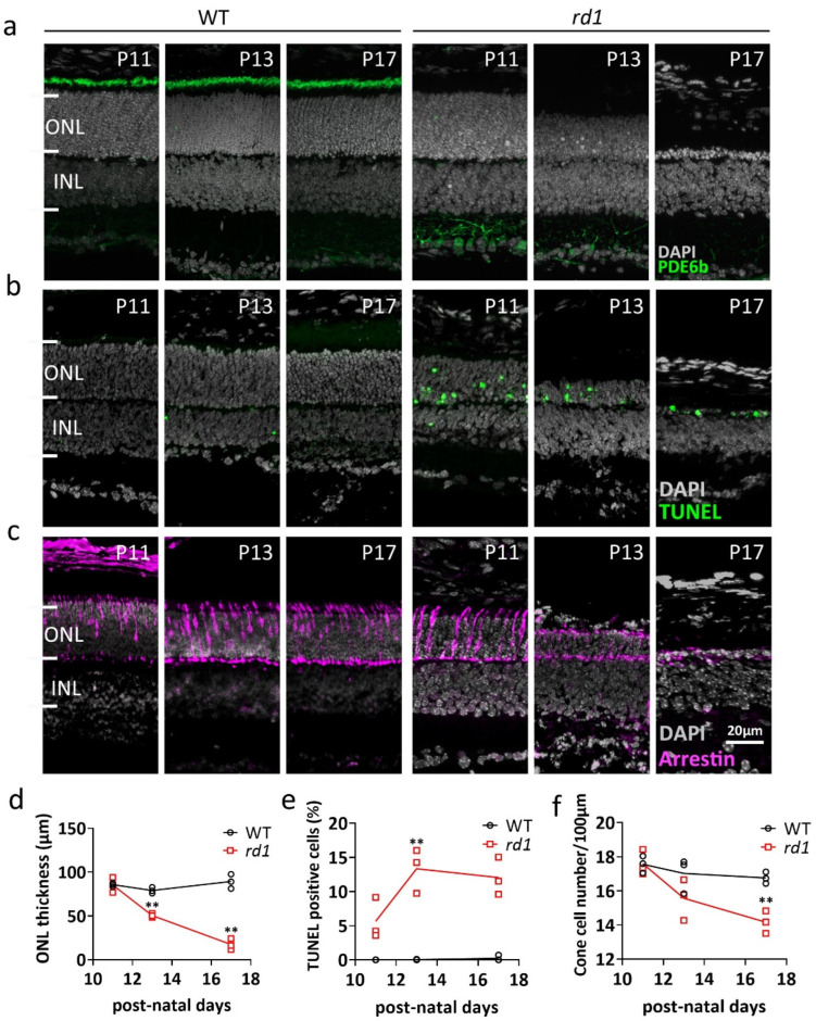
Figure 1.
Comparison of WT and rd1 retina during the 2nd post-natal week. Retinal cross-sections were obtained at postnatal days (P) 11, 13, and 17 from wild-type (WT) and retinal degeneration 1 (rd1) mice. (a) Immunofluorescent staining (IF) for PDE6B (green), labeling photoreceptor outer segments. (b) TUNEL assay (green) showing dying cells in the outer nuclear layer (ONL). (c) IF for cone arrestin (magenta) in the ONL. (d) Quantification of ONL thickness, (e) TUNEL-positive cells, and (f) cone cell number. Data from n = 3 animals per group, expressed as mean ± SD. Statistical significance was assessed using Student’s t-test; significance levels were: ** p < 0.01. DAPI (grey) was employed as nuclear counterstain. INL, inner nuclear layer.

