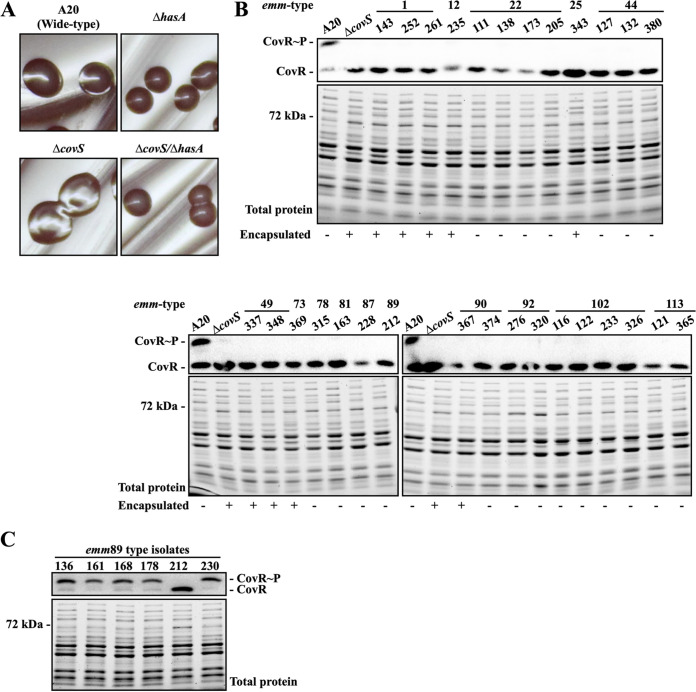
FIG 1.
Colony morphology and expression of phosphorylated CovR in the selected clinical isolates and in the wild-type A20 strain and its covS isogenic mutants. (A) Colony morphology of the wild-type A20 strain, the covS mutant, and their hasA mutants. (B) Expression of phosphorylated CovR and the total protein profile upon SDS-PAGE of selected clinical isolates. (C) Phosphorylation level of CovR in the emm89 isolates. The phosphorylated CovR protein was detected by Phostag Western blotting. Encapsulated colonies are denoted by a plus (+) and unencapsulated colonies are denoted by a minus (−) as shown below the SDS-PAGE gel and Western blot images.

