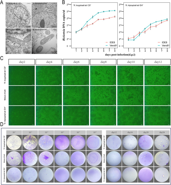
FIG 2.
Comparison of growth characteristics between Rickettsia hoogstraalii str CS and Rickettsia rhipicephalii str EH. (A) Transmission electron micrographs of Vero 81 cells infected with R. hoogstraalii str CS and R. rhipicephalii str EH. Photomicrographs were captured with an H7650 transmission electron microscope camera. (B) Growth curves of R. hoogstraalii str CS and R. rhipicephalii str EH in Vero 81 cells and IDE8 tick cells over 196 h. Error bars represent the standard deviation of the mean. (C) Cytopathic effect in Vero 81 cells induced by R. hoogstraalii str CS, R. rhipicephalii str EH, or a 1:1 mixture of both species (scale bar = 20 μm). (D) Plaque formation in Vero 81 cells by R. hoogstraalii str CS, R. rhipicephalii str EH, or a 1:1 mixture of both species at multiple MOIs and times. (i) Plaque formation with multiple MOIs at 13 days postinfection (dpi). (ii) Plaque formation after inoculation with 2 × 10³ copies/μL at different dpi.

