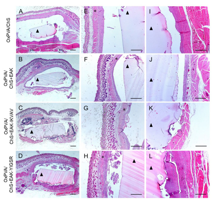
Figure 5.
Hematoxylin and Eosin staining of OxPVA/ChS-based scaffolds integrated with surrounding host tissues at the site of implant (A–D) and preliminary evaluation of the tissues at the superficial (E–H) and deep (I–L) aspects of the grafts. Scale bar: 800 µm (A–H); 400 µm (I–L). (*: the panniculus carnosus within the host subcutaneous tissue; ▲: the OxPVA-based implants).

