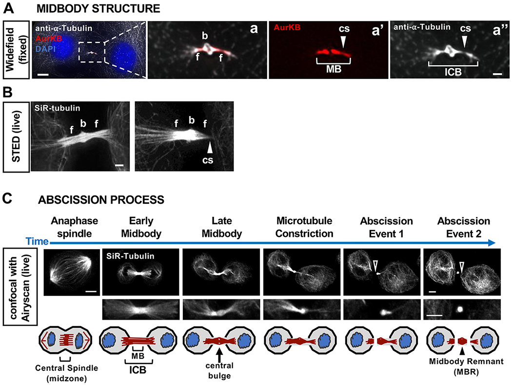
Fig. 2.

Midbody subdomains and abscission process. (A) Widefield image of a mouse embryonic fibroblast at a late stage of abscission, fixed and immunostained for alpha-tubulin (white) and Aurora B kinase (AurKB, red, MB flanks). ICB, intercellular bridge; b, bulge; f, flank; cs, constriction site. (B) The microtubule organization of midbodies is revealed by labeling live HeLa cells with SiR-Tubulin, and stimulated emission depletion (STED) microscopy. (C) Steps in the abscission process visualized in live cells with SiR-tubulin. The midbody is formed by the compaction of central spindle microtubules, then matures, gradually becoming thinner with a central bulge and constriction sites. Microtubules are disassembled to sever each flank (arrowhead), completing the separation of daughter cells and releasing the MBR extracellularly. HeLa cells were imaged by time-lapse confocal microscopy with Airyscan. Scale bars: 1 μm in Aa-Aa”,B; 5 μm in A,C. See [36] for methods
